This research field concerns the investigation of how brain activity is reflected in ear-EEG recordings. It is an ambition to develop a framework for predicting ear-EEG for given cortical activation; and to develop methods, using machine learning methods, that can learn an individual non-parametric forward model, based on EEG recordings, without detailed anatomical information from the individual subject. Moreover, these models will be utilized in the decoding of sensory perception and brain-states.
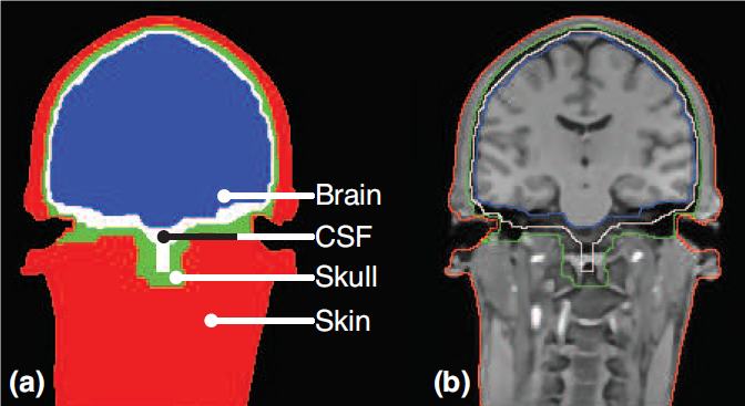
The human head can be regarded as a complex, inhomogeneous, but linear volume conductor (Nunez and Srinivasan, 2006). It follows, that the electric field in the brain and on the surface of the scalp are related to the current from the underlying neural sources through volume conduction. The volume conductor of the head is characterized by the composition of the head and the physical properties of the different tissue types. The conductor can be described in a head model derived from anatomical magnetic resonance imaging (MRI) and is typically composed of four different segments: the brain, the cerebrospinal fluid (CSF), the skull and the scalp (Figure 1).
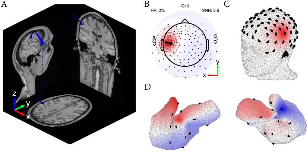
Head models are the basis for computational forward models that describe the transfer function from sources in the brain volume to the electrodes on the surface of the scalp. Forward models can thus be used to estimate the location and direction of an underlying neural source based on high-density EEG scalp recordings (by solving the inverse problem). The forward model can also be used directly to compute the topography of the surface potential of a neural source with known location and direction. At present, methods for estimating forward models for scalp EEG are well-established (Acar and Makeig, 2010), but they offer only a very limited description of the ears. It is an objective to continue our ongoing work to develop and refine specialized ear-EEG forward models based on individual head models that combine MRI scans of the head with scans of ear impressions to obtain high resolution descriptions of the ears. Such models will enable us to predict which cortical phenomena are observable from the ear. Figure 2 illustrates how a single cortical source maps to the scalp and to the ear-canals.
Moreover, forward models can be used to compute the sensitivity distribution to neural sources for a given electrode configuration. This is illustrated in Figure 3 for both a contralateral and an ipsilateral electrode configuration. This provides an intuitive insight into which sources can be measured with ear-EEG, and provide a way to optimize electrode positions in ear-EEG.

The field of neuroscience provides a tremendous knowledge base of how different brain function maps to the cortical structures. One of the aims is to develop a tool that can be used to predict which neurological phenomena can be observed from ear-EEG, and how to place the electrodes in the ear in order to get the best possible sensitivity and specificity.
